【影像征象】靴形心 | The boot-shaped heart sign

欢迎添加小编微信!
.......
Appearance
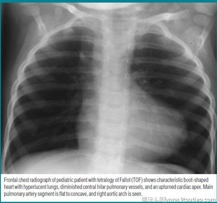
The boot-shaped heart sign is seen on the frontal chest radiograph of children with decreased pulmonary vasculature. The left cardiac border resembles the shape of a wooden boot (Figure) .靴形心见于儿童胸片正位,伴肺血减少。左心缘类似于木靴的形状(图1)。

图1

儿童法洛四联症胸片正位显示特征性靴形心,两肺透亮度增加,中央肺门肺血减少,心尖上翘。肺动脉段凹陷,右位主动脉弓。
Explanation
The boot-shaped heart sign is a conventional radiographic finding in patients with TOF. 靴形心是TOF的常见影像学表现。
The toe of the boot is formed by the upward pointing cardiac apex, which makes an acute angle with the diaphragm.
靴尖由上翘心尖形成,与横膈成锐角。
The upturned cardiac apex is ascribed to right ventricular hypertrophy and occurs in 65% of patients with TOF. 心尖上翘是由于右心室肥厚,见于65%的TOF患者。
The narrower upper part of the boot results from a small or absent main pulmonary artery partly caused by a narrow infundibulum and hypoplastic main pulmonary artery.
靴子上部较窄是由于的肺动脉小或缺失,部分由于漏斗部狭窄和的肺动脉发育不良。
Discussion
The boot-shaped heart is often seen in patients with TOF, which is the most common cause (75%) of cyanotic heart disease.
靴形心最常见于TOF患者,其为发绀性心脏病最常见的原因(75%)。
TOF has four components: (a) obstruction of the right ventricular out flow tract, which may be below the pulmonic valve in the infundibulum of the right ventricle; (b) ventricular septal defect (VSD); (c) overriding of the aorta above the VSD; and (d) right ventricular hypertrophy.
TOF的四个关键点:(a)右心室流出道梗阻,其可能低于右心室漏斗部的肺动脉瓣;(b)室间隔缺损(VSD);(c)VSD上主动脉骑跨;(d)右心室肥大。
Right aortic arch occurs in 20%–30% of patients with cyanotic TOF.
20%~30%的发绀型TOF患者伴位右主动脉弓。
When TOF is also associated with an atrial septal defect, it is called the pentalogy of Fallot.
当TOF也合并房间隔缺损时,称为法洛五联症。
The upturned cardiac apex is usually ascribed to concentric right ventricular hypertrophy.
心尖上翘通常是由于右心室肥厚。
The clinical manifestation and radiographic appearance of TOF depend on the size of the VSD and the severity of the right ventricular outflow tract obstruction; patients become cyanotic at birth when severe pulmonary stenosis or atresia is present.
TOF的临床表现和影像学表现取决于VSD的大小和右室流出道梗阻的严重程度;当出现严重的肺动脉狭窄或闭锁时,患者在出生时就会发绀。
If the right-to-left intracardiac shunt is minimal, the patient will be only mildly cyanotic and may be diagnosed later in life. The chest radiograph may be essentially normal, and the only clue might be the common association of mirror image right aortic arch.
如果右向心分流较小,患者只是轻度发绀,在年龄较大时才可能诊断。胸片可基本正常,唯一的线索可能是常见的右位主动脉弓。

参考文献
1.Haider E A . The boot-shaped heart sign.[J]. Radiology, 2008, 246(1):328-9.
2.Potts W J . Congenital heart disease cyanotic children.[J]. California Medicine, 1953, 78(2):101.